

The BCRF ILC Legacy Project
Nine comprehensively characterized patient-derived organoid models of invasive lobular breast cancer, along with the molecular data, built to provide researchers the faithful lobular models the field has long lacked, and shared openly with the community.
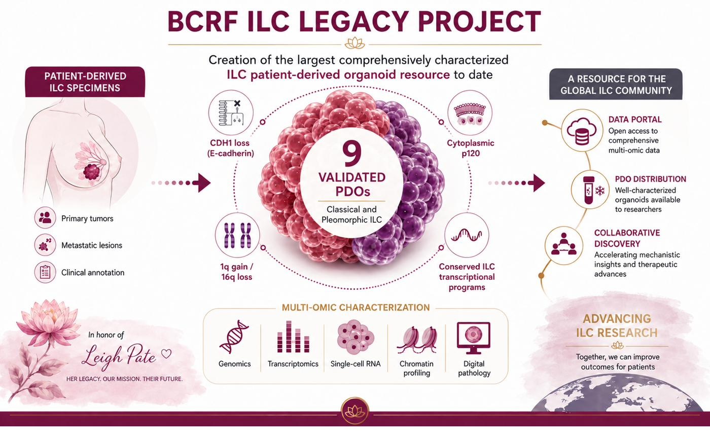
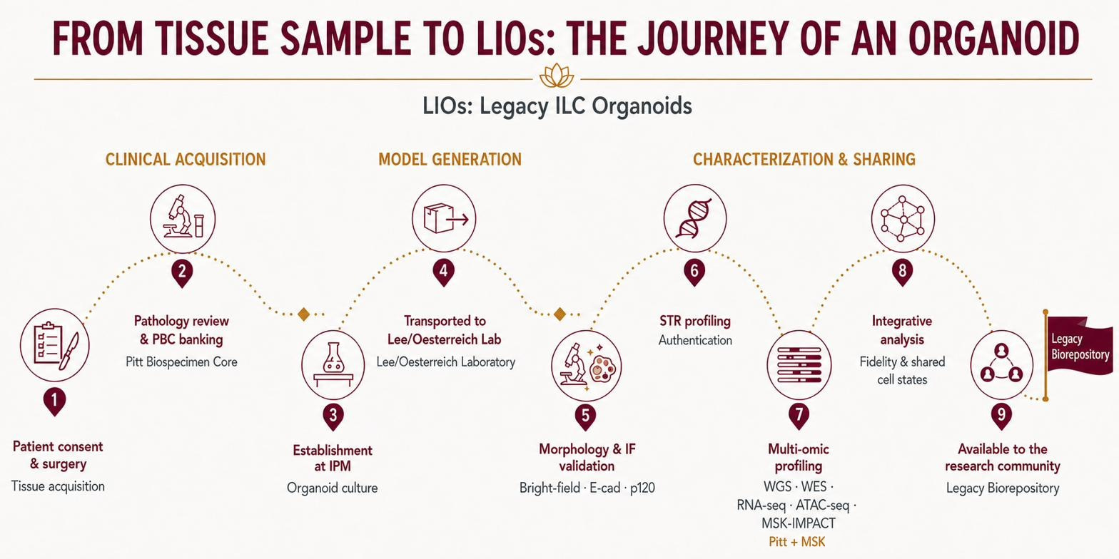
The Legacy collection brings together nine patient-derived organoid lines of invasive lobular breast cancer, each validated against the defining biology of the disease and profiled across up to six molecular layers, alongside data from the very tumor each line came from.

Leigh Pate
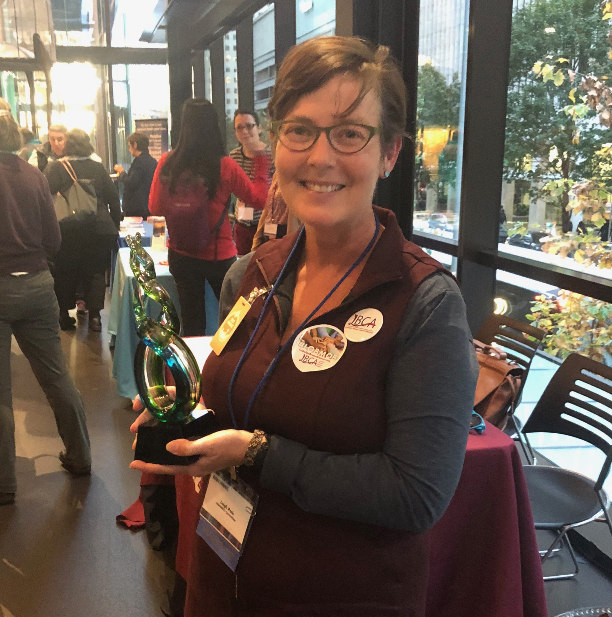
This resource exists because of Leigh Pate. Originally from rural North Carolina and later based in Seattle, Leigh was a writer and communications specialist who, after her 2011 diagnosis with lobular breast cancer, became one of the most influential advocates the ILC community has known.
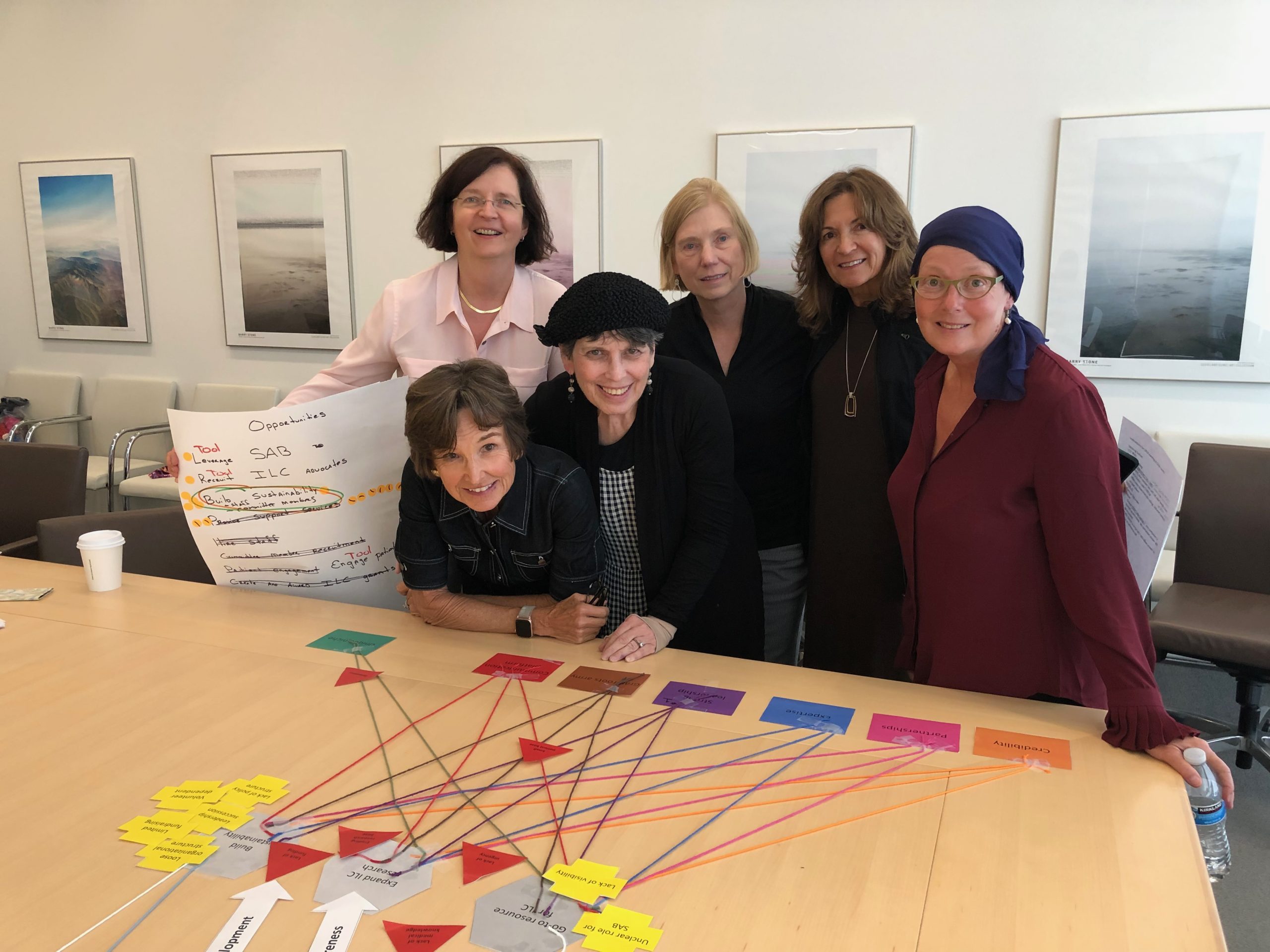
She co-founded the Lobular Breast Cancer Alliance in 2017, shaped its mission, and helped bring lobular breast cancer to the attention of researchers and clinicians worldwide. She led the first white paper defining what was known and unknown about the disease, and was honored as a Susan G. Komen Advocate in Science and Scholar and with the Beth Caldwell Advocacy and Leadership Award.
Though Leigh’s life was cut short in 2022, her gift to the Breast Cancer Research Foundation established this living biorepository and carries her purpose forward. In her honor, the organoid lines are called LIOs, short for Leigh’s Legacy ILC Organoids.

Faithful models for a disease that lacked them
Invasive lobular carcinoma is the most common special histologic subtype of invasive breast cancer, accounting for roughly 10–15% of diagnoses and tens of thousands of new cases each year in the US. Lobular cells lose E-cadherin, the protein that holds cells together, so they grow in single-file strands rather than a lump. That makes invasive lobular carcinoma harder to see on imaging and prone to spreading to unusual places such as the abdomen, ovaries, and gastrointestinal tract.
For decades, invasive lobular carcinoma research has been held back by one gap: too few laboratory models that actually behave like lobular cancer. Leigh Pate understood this, and her gift set out to close it.
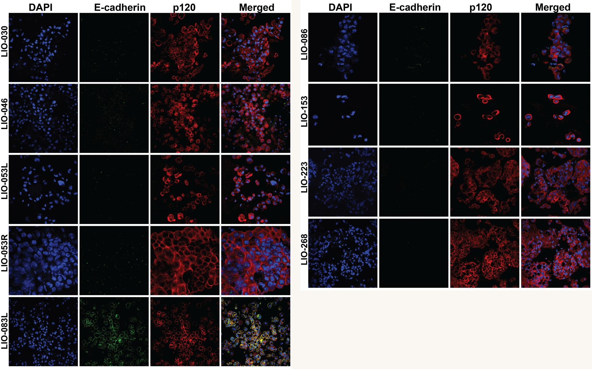
Invasive lobular tumors are slow-growing and notoriously hard to establish in the lab. Building this collection took many attempts, patient growth monitoring, and rigorous validation: every candidate was tested for the molecular hallmarks of lobular cancer, loss of E-cadherin and cytoplasmic p120, and only those that genuinely reproduced that biology were kept. The result is nine validated organoid lines that faithfully carry the biology of the patients they came from.
In total, more than 30 attempts were made to establish ILC organoids from patient tissue. After establishment and growth-phenotype screening, 9 lines were validated by immunofluorescence for the lobular hallmarks, loss of E-cadherin and cytoplasmic p120, to form the final Legacy cohort.
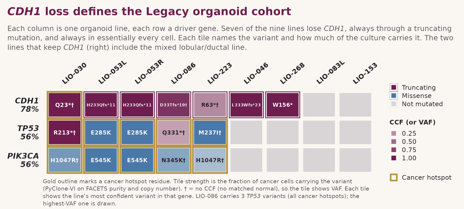
CDH1 loss is the lobular signature, and the organoids keep it
Loss of the CDH1 gene, which makes the cell-adhesion protein E-cadherin, is the molecular hallmark of invasive lobular breast cancer. Seven of the nine Legacy lines carry a CDH1-truncating mutation, faithfully mirroring the disease they model. The two that keep CDH1 intact include the cohort’s one mixed ductal/lobular line, exactly as expected.
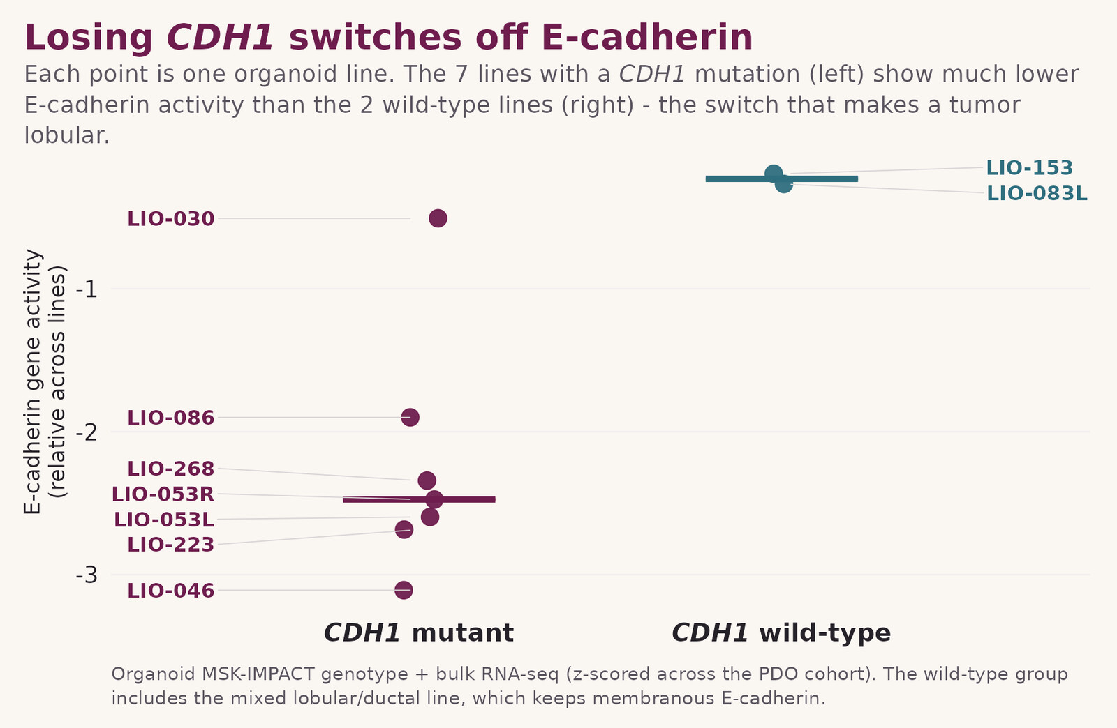
Losing CDH1 switches off E-cadherin, exactly as invasive lobular carcinoma does
The CDH1 mutation doesn’t just sit there. It has the expected biological effect. The seven organoid lines with a CDH1 mutation show sharply lower E-cadherin activity than the two lines that keep CDH1 intact, the molecular switch that defines lobular breast cancer.
Immunofluorescence confirms the lobular phenotype, line by line
Beyond the genetics, every organoid line was directly imaged for the proteins that define lobular cancer. Across the classical and pleomorphic invasive lobular carcinoma lines, E-cadherin is lost from the cell membrane and p120 shifts into the cytoplasm, the same hallmark pattern seen in patient tumors. The cohort’s one mixed ductal/lobular line, LIO-083L, stands apart, retaining clear membranous E-cadherin signal.
The organoids stay true to the patient’s tumor
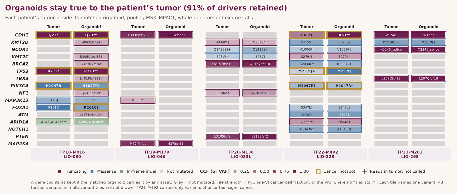
When each organoid is compared with the tumor it came from, the same cancer-driving mutations show up in both. Across five matched patient–organoid pairs, 91% of the tumor’s driver genes are retained in the organoid, including CDH1, in every single case. These models aren’t just similar to the original cancer; they’re faithful working copies of it.
A shared landscape of cell states across patients
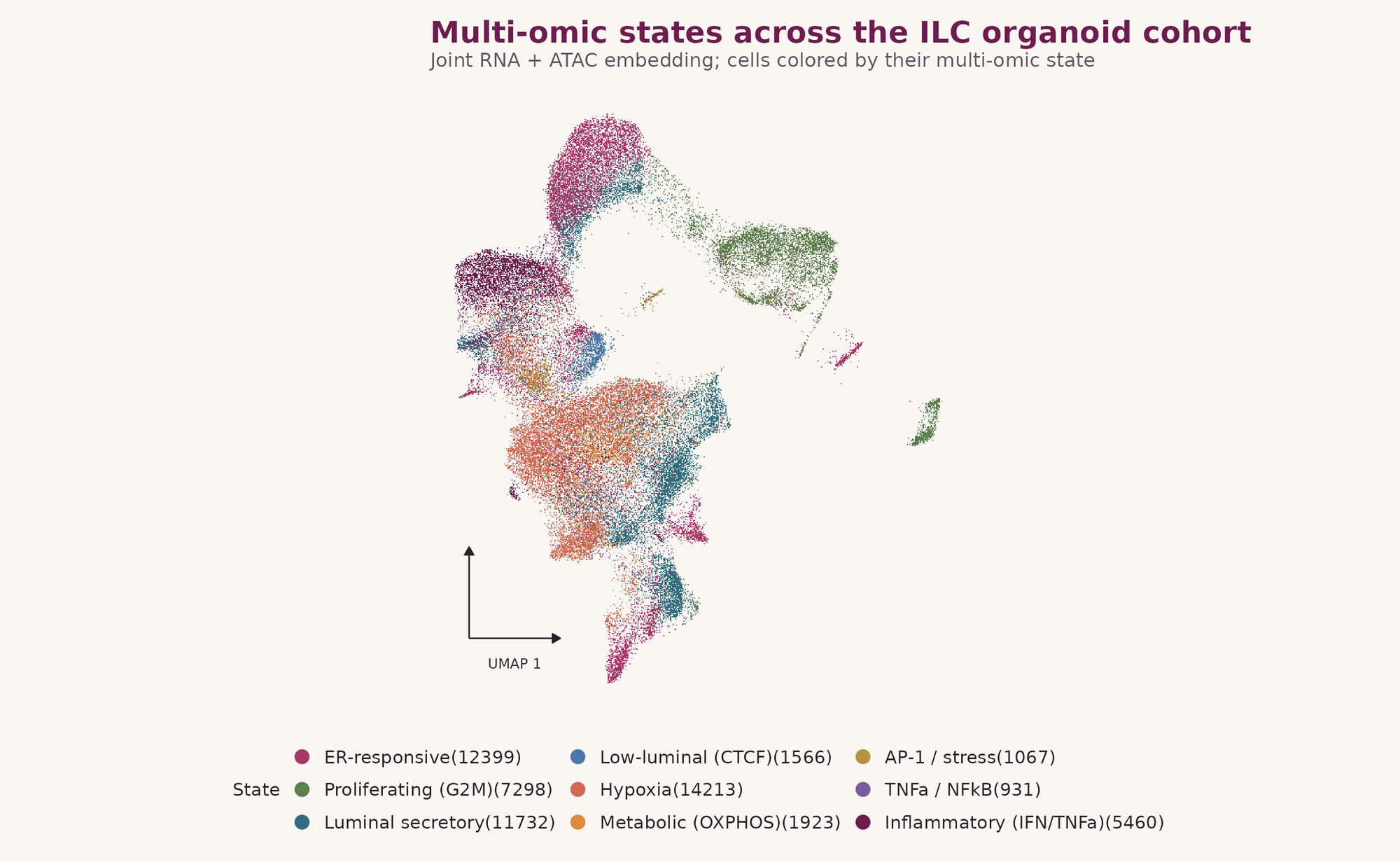
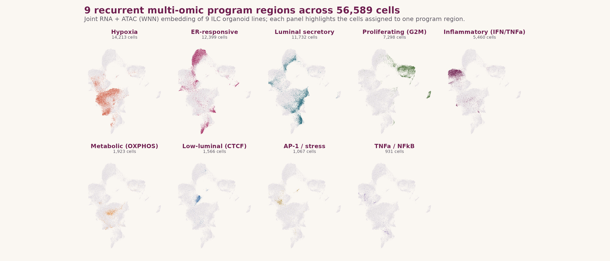
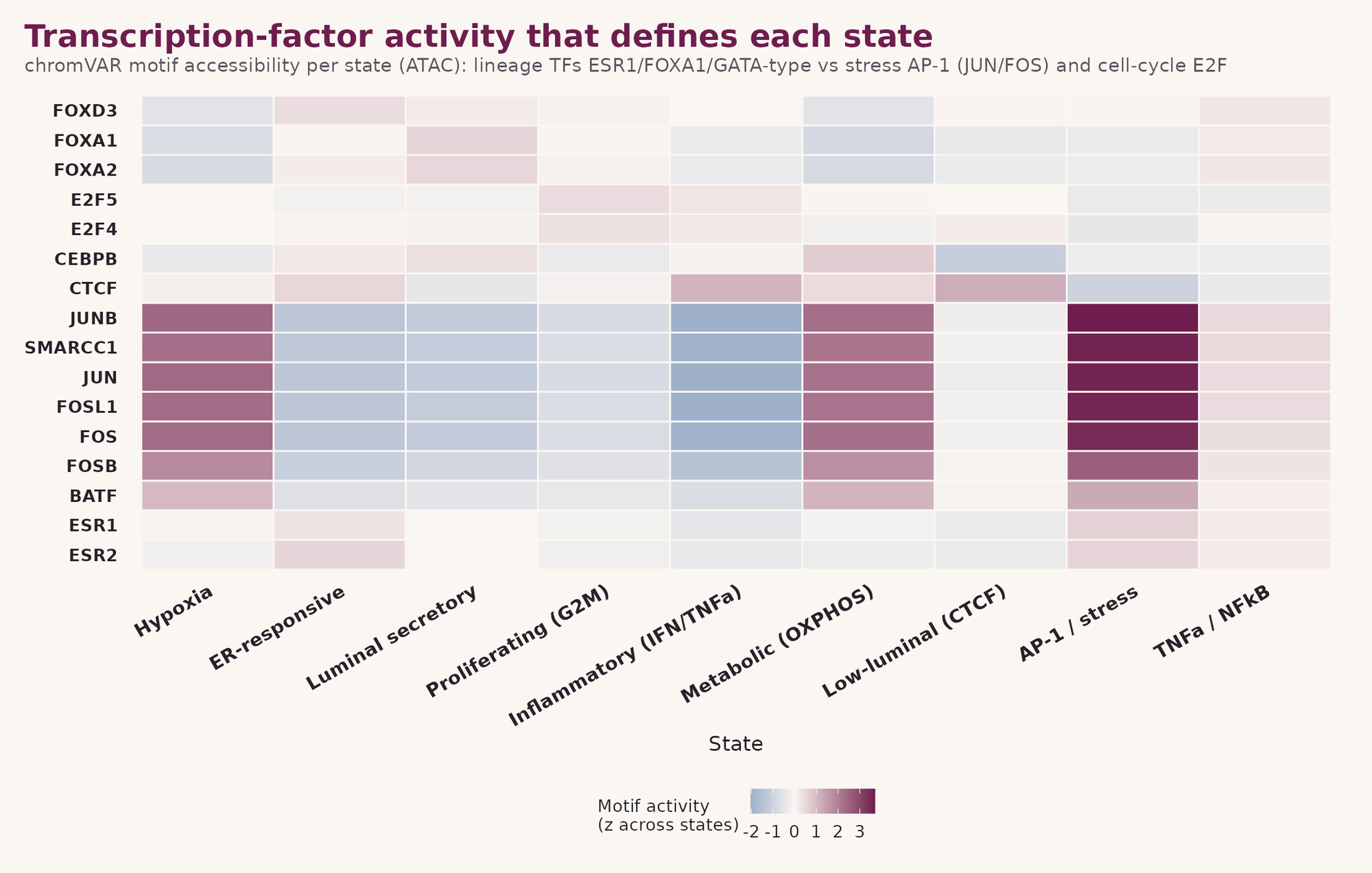
Looking at the organoids one cell at a time, using combined gene-expression and chromatin-accessibility profiling across more than 56,000 cells, cells settle into nine recurring molecular states that reappear across different patients, suggesting they reflect core biology of invasive lobular cancer.
Explore the state-level data
The full multi-omic picture behind the summary above, for researchers who want to go deeper into how these states were defined, where they sit across the cohort, and what drives them. Click any figure to enlarge.
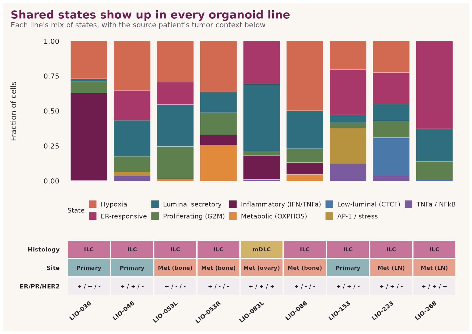
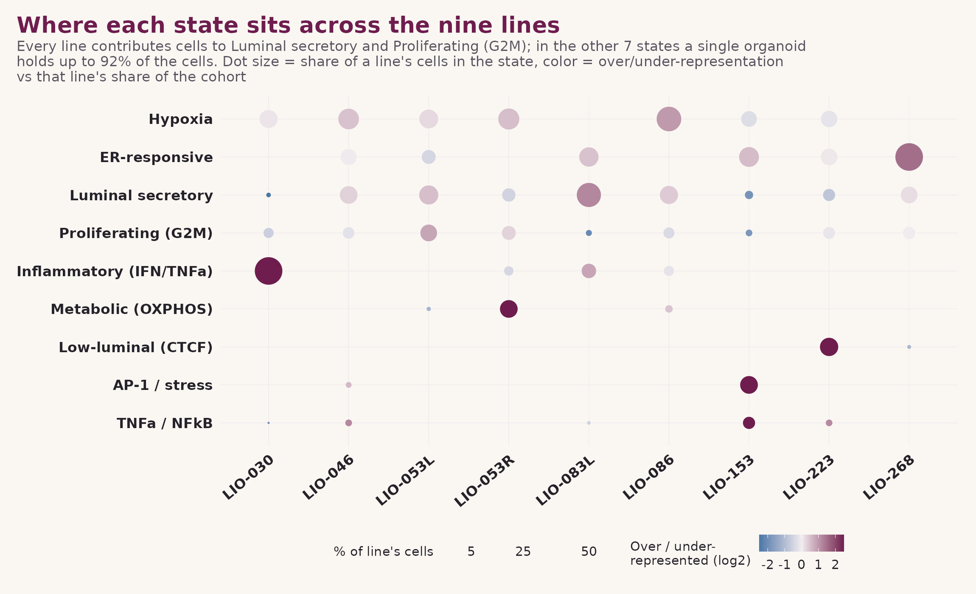
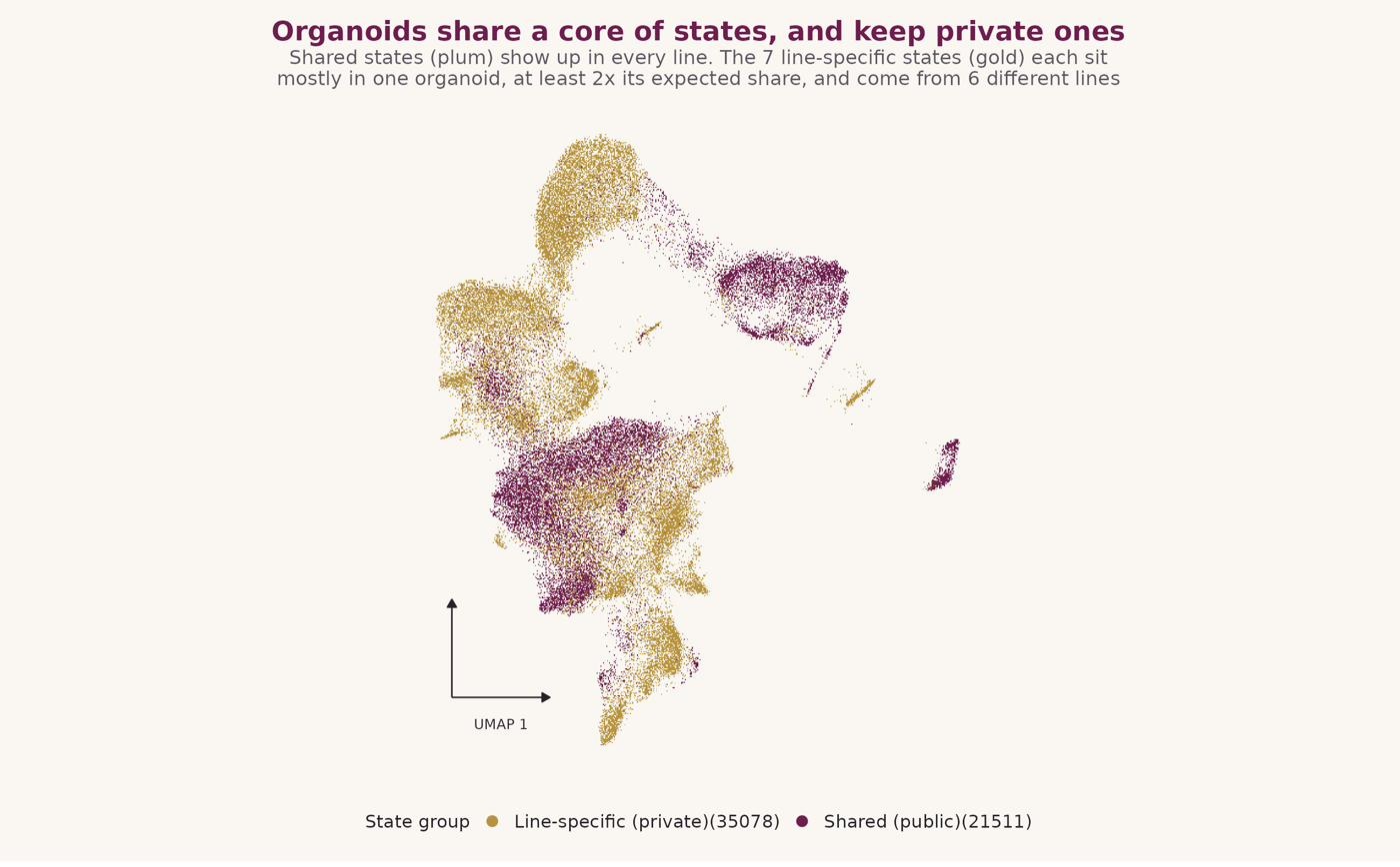
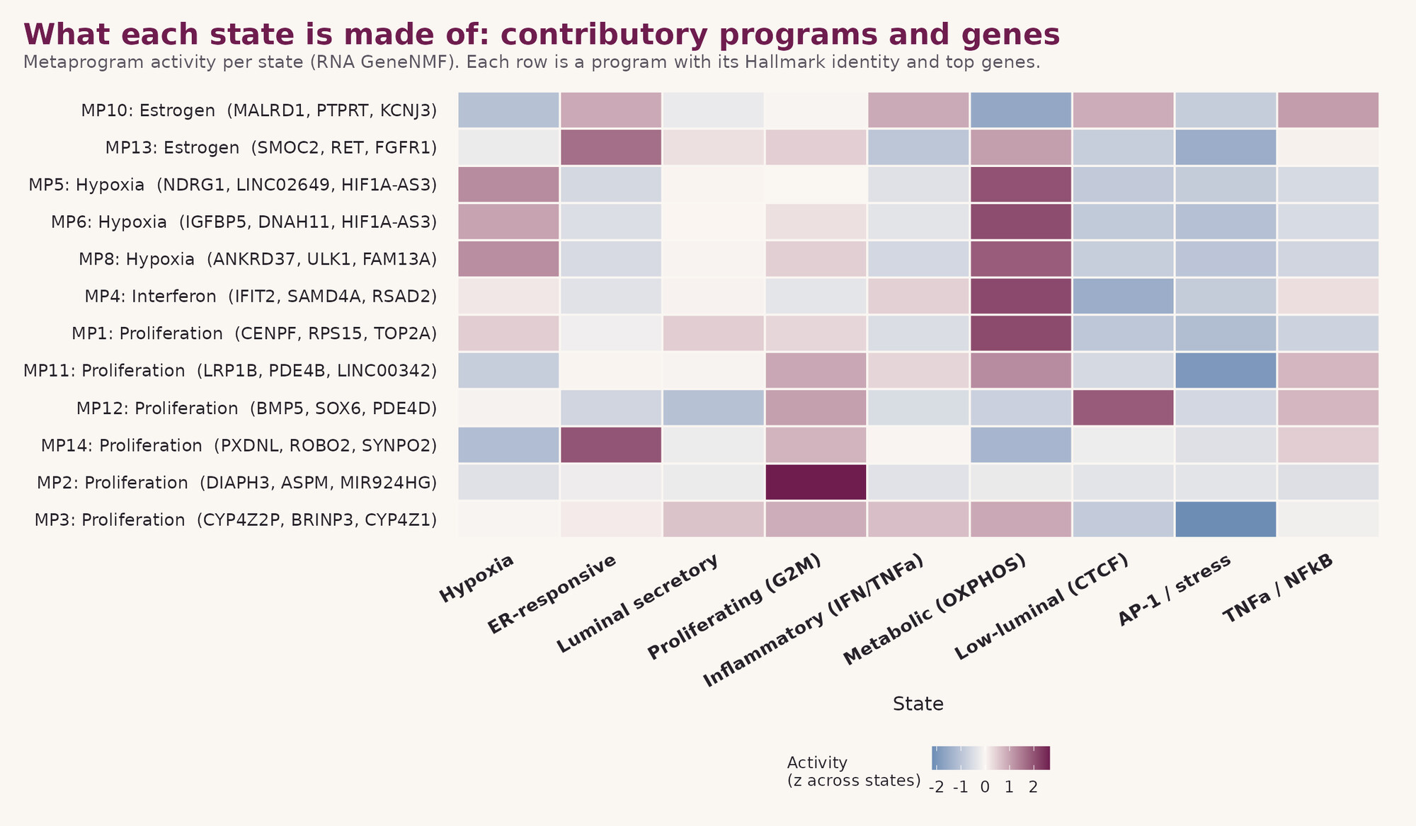
The Legacy organoid lines
All nine lines at a glance. Each line is accompanied by its full molecular dataset; matched patient-tumor data (whole-genome sequencing, digital pathology) is available for a subset.
| Line | Histology | Origin | ER / PR / HER2 | E-cad / p120 | Data available (organoid) |
|---|---|---|---|---|---|
| LIO-030* | ILC | Primary | + / + / − | − / cyto | WES · bulk RNA-seq · snRNA-seq · snATAC-seq · MSK-IMPACT · IF |
| LIO-046 | ILC (pILC) | Primary | + / + / − | − / cyto | WES · bulk RNA-seq · snRNA-seq · snATAC-seq · MSK-IMPACT · IF |
| LIO-053L | ILC | Metastasis, bone | + / − / − | − / cyto | WES · bulk RNA-seq · snRNA-seq · snATAC-seq · MSK-IMPACT · IF |
| LIO-053R | ILC | Metastasis, bone | + / − / − | − / cyto | WES · bulk RNA-seq · snRNA-seq · snATAC-seq · MSK-IMPACT · IF |
| LIO-086 | ILC | Metastasis, bone | + / − / − | − / cyto | WES · bulk RNA-seq · snRNA-seq · snATAC-seq · MSK-IMPACT · IF |
| LIO-153 | ILC | Primary | + / + / − | − / cyto | WES · bulk RNA-seq · snRNA-seq · snATAC-seq · MSK-IMPACT · IF |
| LIO-223 | ILC | Metastasis, lymph node | + / + / − | − / cyto | WES · bulk RNA-seq · snRNA-seq · snATAC-seq · MSK-IMPACT · IF |
| LIO-268 | ILC (pILC) | Metastasis, lymph node | + / + / + | − / cyto | WES · bulk RNA-seq · snRNA-seq · snATAC-seq · MSK-IMPACT · IF |
| LIO-083L* | mixed (mDLC) | Metastasis, ovary | + / + / + | + / mem | WES · bulk RNA-seq · snRNA-seq · snATAC-seq · MSK-IMPACT · IF |
Access the models and data
Request the organoids
All nine Legacy organoid lines are available to the research community through the Organoid Research Core (ORC) at the University of Pittsburgh.
Request a line →Explore the data
A dedicated data portal with full access and visualization tools is in development. This section will be updated with details on access and visualization as they become available.
The people and partners behind the Legacy Project
The Legacy project is led by the Lee/Oesterreich Laboratory at the University of Pittsburgh / UPMC Hillman Cancer Center and Magee-Womens Research Institute, in partnership with Memorial Sloan Kettering Cancer Center and the Institute for Precision Medicine, and supported by the Breast Cancer Research Foundation in honor of Leigh Pate.
University of Pittsburgh
-
Adrian LeePhDWomen’s Cancer Research Center, UPMC Hillman Cancer CenterInstitute for Precision Medicine, University of PittsburghDepartment of Pharmacology & Chemical Biology, University of Pittsburgh, Pittsburgh, PA, USA
-
Brent T. SchlegelBSLee/Oesterreich Laboratory, University of Pittsburgh, Pittsburgh, PA, USA (computational analysis)Department of Pharmacology & Chemical Biology, University of Pittsburgh, Pittsburgh, PA, USA
-
Daniel D. BrownPhDInstitute for Precision Medicine, University of Pittsburgh, Pittsburgh, PA, USA
-
Jagmohan HoodaPhD, MBAWomen’s Cancer Research Center, University of Pittsburgh Medical Center (UPMC), Hillman Cancer CenterMagee-Womens Research Institute, Pittsburgh, PA, USA
-
Jian ChenMSWomen’s Cancer Research Center, University of Pittsburgh Medical Center (UPMC), Hillman Cancer CenterMagee-Womens Research Institute, Pittsburgh, PA, USA
-
Priscilla F. McAuliffeMDDepartment of Breast Surgical Oncology, University of Pittsburgh, Pittsburgh, PA, USA
-
Rohit BhargavaMDDepartment of Pathology, University of Pittsburgh, Pittsburgh, PA, USA
-
Steffi OesterreichPhDWomen’s Cancer Research Center, UPMC Hillman Cancer CenterDepartment of Pharmacology & Chemical Biology, University of Pittsburgh, Pittsburgh, PA, USA
Memorial Sloan Kettering Cancer Center
-
Britta WeigeltPhDDepartment of Pathology and Laboratory Medicine, Memorial Sloan Kettering Cancer Center, New York, NY, USA
-
Fresia ParejaMD, PhDDepartment of Pathology and Laboratory Medicine, Memorial Sloan Kettering Cancer Center, New York, NY, USA
-
Pier SelenicaMSDepartment of Pathology and Laboratory Medicine, Memorial Sloan Kettering Cancer Center, New York, NY, USA
-
Hunter GreenBSDepartment of Pathology and Laboratory Medicine, Memorial Sloan Kettering Cancer Center, New York, NY, USA
-
Kaitlyn GillBSDepartment of Pathology and Laboratory Medicine, Memorial Sloan Kettering Cancer Center, New York, NY, USA
-
Jorge Reis-FilhoMD, PhD
Conference Presentations
- Metastasis Research Society (MRS) 21st Biennial Congress, 2026 (Poster Link)
- MCBIOS Annual Conference, 2026 (Poster Link)
- San Antonio Breast Cancer Symposium (SABCS), 2024 (Poster Link)
- International ILC Symposium, 2024 (Poster Link)
With Thanks To