

Dr. Steffi Oesterreich and Leigh Pate became good friends (and Leigh became a fierce yet humble ILC patient advocate). They had many phone calls and some in-person meetings discussing how to drive ILC research forward. Other patients who had joined the 1st ILC Symposium in 2016 in Pittsburgh joined in (such as Lori Pettiti) – and the Lobular Breast Cancer Alliance (LBCA) was born. Dr. Steffi Oesterreich served as Chair of the Scientific Advisory Board until 2022.

An unbiased screen performed in Dr. Adrian Lee’s lab, identified E-cadherin as a top candidate interacting with the IGF1 pathway. Dr. Lee joined the ILC research efforts, which propelled ILC genomic studies in the now combined Lee-Oesterreich lab.
After many discussions and guidance from Dr. Davidson, we opened the first clinical trial specifically in ILC: a biomarker window trial aimed at improving understanding of endocrine therapy in patients with ILC. It was performed with the help of the Translational Breast Cancer Research Consortium (TBCRC). Drs. Rachel Jankowitz and Priscilla McAuliffe are clinical PIs. Dr. Jankowitz received a Career Development Award from Susan G. Komen supporting the study.
Here is the press release: https://www.upmc.com/media/news/100615-invasive-lobular-breast-cancer

After some unsuccessful attempts to obtain NIH funding, Dr. Oesterreich received BCRF funding for her ILC research. This allowed the journey to start. We increased research on ILC, with the support of some exceptional students, postdocs and Dr. Nancy Davidson, at the time Dr. Oesterreich’s main mentor. Of note BCRF also provided funds for subsequent ILC Symposia, and started to include ILC into Breakout sessions at the Annual BCRF Symposium.

When moving from Baylor College of Medicine, Houston, TX, to UPCI/Magee Womens Research Institute, Dr. Oesterreich had just become interested in ILC, as some estrogen receptor cofactors were expressed higher in ER+ ILC (as per Oncomine). She decided to start researching ILC, and was delighted to learn that Dr. David Dabbs, the Chief Pathologist at UPMC Magee-Womens Hospital was an internationally recognized expert in ILC. The ILC studies would not have been possible without Dr. Dabbs advice and countless slide reviews. Dr. Oesterreich and Dr. Dabbs had many productive collaborations, and also co-authored a review in “Breast Pathology, 2nd Edition by David J. Dabbs”.

Dr. Oesterreich gives a Keynote Lecture at the ILC Course in Boston in 2018, which starts the official continuation of the ILC Symposia. Drs. Otto Metzger and Rachel Jankowitz worked with some amazing advocates, including Julia Levine, Janice Axelroad, Lori Pettiti and Flora Migyanka (who later founded Dynami)
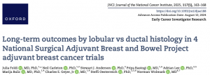
In this study, we performed a combined analysis of four NSABP trials with more than 12,000 patients treated with anthracycline-based adjuvant chemotherapy, and with at least 10 years of follow-up. While there was no significant difference in disease free survival and overall survival over the entire follow-up period, stratification by years since diagnosis showed that patients with ILC exhibited increased risk for late DFS events, recurrences, and deaths. We are currently expanding our correlative studies using the primary tumor specimens from this unique, large, annotated cohort of patients with ILC and matched NST with long-term follow up.
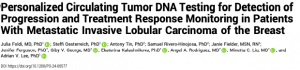
In a collaboration with Natera, we analyzed ctDNA longitudinally in 66 patients with metastatic ILC using tumor-informed Signatera assays. Sequencing results from N=355 samples showed that ctDNA testing is a promising approach for surveillance and precision medicine in patients with metastatic ILC, a setting where imaging has often limited sensitivity.
It remains challenging to understand and categorize mixed ductal lobular tumors. In this study, we utilized spatially resolved transcriptomic, genomic, and single-cell profiling for a comprehensive characterization of three mixed cases (categorized as such by three expert breast cancer pathologists at UPMC, UM and MSKCC). We identified clinically significant differences between ductal and lobular tumor regions including distinct intrinsic subtype heterogeneity – a finding that indicates challenges for prognosis and therapeutic decisions for these cases.
Together with patient advocates and leading ILC experts, we designed a survey in which with more than 1,700 (researchers, clinicians and patients) from 66 countries participated. Patients identified improvement of ILC screening/early detection, and development of better imaging tools as top research priorities. In contrast, both researchers and clinicians identified understanding of endocrine resistance and identifying novel drugs that can be tested in clinical trials as top research priority. Other research priorities included i) Dormancy, ii) ILC modeling, and ii) genomic predictors. Other noteworthy findings were that the majority of patients and advocates thought that their health care providers did not sufficiently explain the unique features of ILC, and that 85% of clinicians have never powered a clinical trial to allow subset analysis for histological subtypes, but the majority would consider it, and would participate in an ILC clinical trials consortium.